Mouse Model for Pulmonary Embolism (PE) ")

- UOM
- FOB US$ 140.00
- Quantity
Overview
Properties
- Product No.DSI517Mu01
- Organism SpeciesMus musculus (Mouse) Same name, Different species.
- ApplicationsDisease Model
Research use only - Downloadn/a
- Category
- Prototype SpeciesHuman
- SourceThrombin induced
- Model Animal StrainsBalb/c mice (SPF class), male, weeks in age:4weeks~6weeks, 20g~22g.
- Modeling GroupingRandomly divided into groups: Control group, Model group, Positive drug group and Test drug group, 15 mice per group.
- Modeling Period1~2 days
Sign into your account
Share a new citation as an author
Upload your experimental result
Review
Contact us
Please fill in the blank.
-
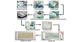
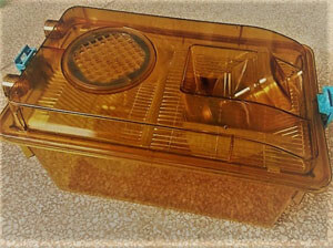 Packages (Simulation)
-
 Packages (Simulation)
-
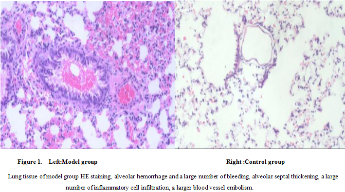 Fig. HE staining of lung in model group and control group
-
 ISO9001: 2008, ISO13485: 2003 Registered
Modeling Method
1. 22g-26g mice, with 3% Pentobarbital Sodium, with 80mg/kg intraperitoneally anesthetized mice, mice in supine position, the limbs and head with tape fixed on anatomical plate, with 8% sodium sulfide to remove the neck hair, povidone iodine disinfection in preparation for surgery.
2. Incision on the middle of the neck, the separation of soft tissue and muscle, exposure to the left side of the jugular vein, inject
Thrombin solution (soluble in 0.9% normal saline, dose 20~75U/kg) to the internal jugular vein .
3. Clean up the operation field, suture the neck incision, stay awake and put mice back to clean cage, observe the state and the state of the mouse and record.
4. Sham operation is the same as that operation in modeling group,but the normal saline injected into the jugular vein.
5. After 24hrs, kill the mice, take the left lung tissue, 4% poly formaldehyde solution fixed, paraffin embedded; the rest of the lung tissue -80 degree preservation.
Model evaluation
Peripheral blood platelet count:
collect 20ul mice peripheral blood , put in 380 μL (10g/L) ammonium oxalate dilution, after mixing and use blood cell counting plate platelet count.
Pathological results
Take the left lung, 4% poly formaldehyde fixed, and then dehydration, paraffin embedded, sliced (thickness of 5μm), HE staining.
A few hours after that alveolar exudation and hemorrhage, alveolar septal thickening, which a large number of inflammatory cell infiltration, visible large artery embolism, platelets and red blood cell aggregation and adhesion to around the embolus.
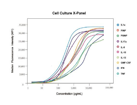
Cytokines level
Statistical analysis
SPSS software is used for statistical analysis, measurement data to mean ± standard deviation (x ±s), using t test and single factor analysis of variance for group comparison , P<0.05 indicates there was a significant difference, P<0.01 indicates there are very significant differences.
Giveaways
Increment services
-
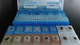 Tissue/Sections Customized Service
-
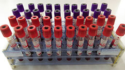 Serums Customized Service
-
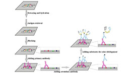 Immunohistochemistry (IHC) Experiment Service
-
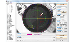
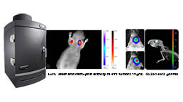 Small Animal In Vivo Imaging Experiment Service
-
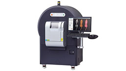 Small Animal Micro CT Imaging Experiment Service
-
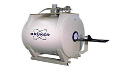 Small Animal MRI Imaging Experiment Service
-
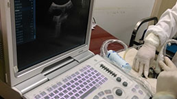 Small Animal Ultrasound Imaging Experiment Service
-
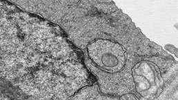 Transmission Electron Microscopy (TEM) Experiment Service
-
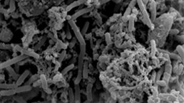 Scanning Electron Microscope (SEM) Experiment Service
-
 Learning and Memory Behavioral Experiment Service
-
 Anxiety and Depression Behavioral Experiment Service
-
 Drug Addiction Behavioral Experiment Service
-
 Pain Behavioral Experiment Service
-
 Neuropsychiatric Disorder Behavioral Experiment Service
-
 Fatigue Behavioral Experiment Service
-
 Nitric Oxide Assay Kit (A012)
-
 Nitric Oxide Assay Kit (A013-2)
-
 Total Anti-Oxidative Capability Assay Kit(A015-2)
-
 Total Anti-Oxidative Capability Assay Kit (A015-1)
-
 Superoxide Dismutase Assay Kit
-
 Fructose Assay Kit (A085)
-
 Citric Acid Assay Kit (A128 )
-
 Catalase Assay Kit
-
 Malondialdehyde Assay Kit
-
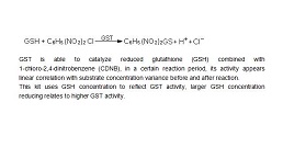 Glutathione S-Transferase Assay Kit
-
 Microscale Reduced Glutathione assay kit
-
 Glutathione Reductase Activity Coefficient Assay Kit
-
 Angiotensin Converting Enzyme Kit
-
 Glutathione Peroxidase (GSH-PX) Assay Kit
-
 Cloud-Clone Multiplex assay kits