- Featured-Product
- SPF
Rat Model for Osteoarthritis (OA) ")

Ostarthritis; Ostearthritis; OA
- UOM
- FOB US$ 280.00
- Quantity
Overview
Properties
- Product No.DSI587Ra04
- Organism SpeciesRattus norvegicus (Rat) Same name, Different species.
- ApplicationsDisease Model
Research use only - Downloadn/a
- CategorySkeletal, articular and cutaneous systems
- Prototype SpeciesHuman
- SourceInduced by surgical resection of anterior and posterior cruciate ligament and medial meniscus
- Model Animal StrainsWistar Rats(SPF), healthy, male, 3-4W, body weight 180g~200g.
- Modeling GroupingRandomly divided into six group: Control group, Model group, Positive drug group and three Test drug group.
- Modeling Period4-6 weeks
Sign into your account
Share a new citation as an author
Upload your experimental result
Review

Contact us
Please fill in the blank.
-
 Packages (Simulation)
-
 Packages (Simulation)
-
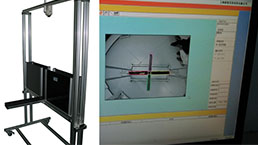
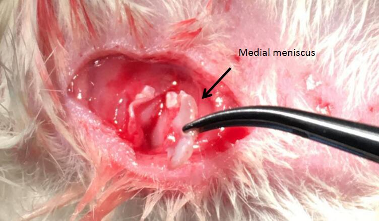 Fig 1. Surgical resection of anterior and posterior cruciate ligament and medial meniscus
-
 ISO9001: 2008, ISO13485: 2003 Registered
Modeling Method
1. Anesthetized with intraperitoneal injection 1% pentobarbital sodium (0.4ml/100g). The right knee joint was shaved and disinfectant with iodine disinfection.
2. Make a incision about 0.5cm long on the right knee medial longitudinal, cut the skin, subcutaneous tissue separation, exposed and cut off the medial collateral ligament, open the knee joint cavity, exposed and cut off the anterior cruciate ligament, exposure and resection of medial meniscus, hemostasis, repeatedly flushing articular cavity with saline and sutured, put Chloramphenicol on the wound to prevent infection.
3. Observe the changes of wound and other postoperative complications in rats,and normally bred after operation for one month. The rats were sacrificed and the knee and hip joints were removed. The knee and hip joint specimens were immersed in 4% paraformaldehyde in 1 days, on the 14% of two sodium EDTA decalcified for 6 weeks, and then tissue embedding and paraffin sections were then performed.
Model evaluation
Pathological results
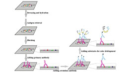
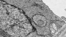
1. Morphological observation of articular cartilage by HE staining:
The normal control group can be seen in the cartilage surface, transitional layer, radiation layer, each layer of calcified layer structure is clear, smooth articular surface, cells arranged in neat and uniform cytoplasm, cartilage surface was pink, with blue nuclei, each layer of uniform dyeing, tide line visible; model group showed that the articular surface is not smooth that is not the whole surface roughness, even spalling, cartilage destruction, cartilage layers are not clear, the visible portion of empty cells arranged in disorder
2. Morphological observation, toluidine blue staining:
In the normal control group, the surface layer, the transfer layer, the radiation layer and the calcified layer of the cartilage can be seen
Clear, smooth articular surface, cells arranged in uniform, each layer of uniform dyeing, the cartilage layer and the nucleus is deep blue,
The tide line is clear; the model group joint cartilage surface is not smooth, the hierarchical structure is not clear, the blue part of the cartilage cells disappeared, uneven staining, the articular cartilage surface, the number of cartilage cells decreased, can see a lot of hypertrophic chondrocytes, tufted cells arranged densely distributed, and irregular tide line is not clear.
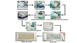
Cytokines level
The contents of IL-1, IL-6 and TNF- alpha in synovial fluid were assayed by ELISA Kit
Statistical analysis
SPSS software is used for statistical analysis, measurement data to mean ± standard deviation (x ±s), using t test and single factor analysis of variance for group comparison , P<0.05 indicates there was a significant difference, P<0.01 indicates there are very significant differences.
Giveaways
Increment services
-
 Tissue/Sections Customized Service
-
 Serums Customized Service
-
 Immunohistochemistry (IHC) Experiment Service
-
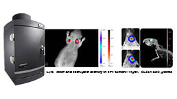 Small Animal In Vivo Imaging Experiment Service
-
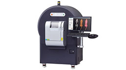 Small Animal Micro CT Imaging Experiment Service
-
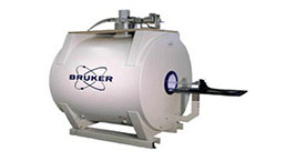 Small Animal MRI Imaging Experiment Service
-
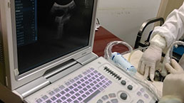 Small Animal Ultrasound Imaging Experiment Service
-
 Transmission Electron Microscopy (TEM) Experiment Service
-
 Scanning Electron Microscope (SEM) Experiment Service
-
 Learning and Memory Behavioral Experiment Service
-
 Anxiety and Depression Behavioral Experiment Service
-
 Drug Addiction Behavioral Experiment Service
-
 Pain Behavioral Experiment Service
-
 Neuropsychiatric Disorder Behavioral Experiment Service
-
 Fatigue Behavioral Experiment Service
-
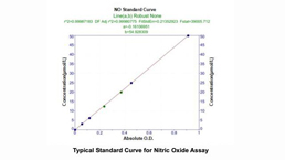
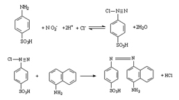 Nitric Oxide Assay Kit (A012)
-
 Nitric Oxide Assay Kit (A013-2)
-
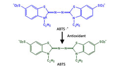 Total Anti-Oxidative Capability Assay Kit(A015-2)
-
 Total Anti-Oxidative Capability Assay Kit (A015-1)
-
 Superoxide Dismutase Assay Kit
-
 Fructose Assay Kit (A085)
-
 Citric Acid Assay Kit (A128 )
-
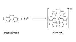
 Catalase Assay Kit
-
 Malondialdehyde Assay Kit
-
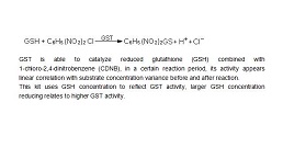 Glutathione S-Transferase Assay Kit
-
 Microscale Reduced Glutathione assay kit
-
 Glutathione Reductase Activity Coefficient Assay Kit
-
 Angiotensin Converting Enzyme Kit
-
 Glutathione Peroxidase (GSH-PX) Assay Kit
-
 Cloud-Clone Multiplex assay kits